| Catheter Ablation | ||||||
 |
Waiting List
|
Hospital Entry
|
Location /Contact
|
|||
|
3 Months
|
Hospital Admission : Same day as op Hospital Stay: 2 Nights Length of Procedure: 3 hours |
Contact: Win Bell |
||||
A.V. Nodal Ablation
In this page : Wolff-Parkinson-White syndrome, supraventricular tachycardia, ventricular tachycardia, atrial fibrillation, atrial flutter, Pacemaker, AV Nodal Ablation, Procedure, Pacemaker insertion, After procedure
Description This page describes AV Nodal ablation. The ablation procedure can be used to treat other conditions which may not require a pacemaker to be inserted such as Wolff-Parkinson-White syndrome. A special catheter is guided up into the heart via an insertion point in the groin. The head of the catheter has electrical sensors which help the cardiologist to guide the catheter to the correct place. Once in place the cardiologist can deliver electrical pulses which destroy (ablate) the tissue which is causing the heart problem.
What is the treatment for : Wolff-Parkinson-White syndrome, supraventricular tachycardia, ventricular tachycardia, atrial fibrillation or atrial flutter.
Specialists : Medical : Dr A. Fitzpatrick, Dr S. Petkar, Dr D. Fox - Nursing: Sister W. Bell
|
Atrial Fibrillation : In this condition the regular pumping function of the upper chambers [the atria] of the heart is replaced by a disorganized, ineffective quivering caused by chaotic conduction of electrical signals through the upper chambers. When the atria contract, an electrical impulse is sent to the lower chambers of the heart [the ventricles], signaling them to contract too. Each impulse passes through the atrio-ventricular [A.V.] node, which acts to regulate the flow of these electrical impulses. In atrial fibrillation this A.V. node tries to cope with the extra electrical impulses but is unable to control them all resulting in the lower chambers also beating in a fast chaotic manner. An Atrioventricular (A.V.) Nodal Ablation. In order to stop these unpleasant symptoms a procedure known as an ablation can be performed. In this procedure the doctor will block [ablate] the A.V. node. By doing this he will remove the connection between the upper and lower chambers.
This is sometimes done deliberately to stop rapid impulses being conducted to the ventricles from the atria through the AV-node. If it is done deliberately, then a pacemaker is always needed. Increasingly, the sophistication of catheter ablation, especially for the treatment of atrial fibrillation, means that we don't need to rely on this relatively simple approach.
Very rarely, a pacemaker is needed when a catheter ablation is done, and the cells of the AV-node are damaged accidently. Usually this happens when a patient is having ablation for SVT, due to AV-nodal re-entry. This accidental damage occurs in about 1:150 cases.
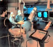
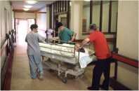
If successful, the ventricles will be unable to respond to the signals transmitted from the atria. If the patient is on tablets to thin the blood [anticoagulants], they will still need to be taken . If tablets to control heart beat are taken they may no longer be needed. As the pathway between the upper and lower chambers of the heart is destroyed during an ablation it is necessary to provide another way of providing the lower chambers with the electrical stimuli they require to make them pump. This can be done by implanting a pacemaker. The pacemaker consists of a small box [or pulse generator], attached to one or two leads [or wires] which are passed into the heart. The box is about the size of a matchbox and weighs approximately 50-100g [2-4oz]. It has an outer casing of metal which is sealed. Inside the box is a battery and an electronic circuit. The wires are introduced into the main vein on the left side of the neck and positioned in the heart. Once in position they are secured and the box is attached. The box is then placed in a small pocket created between the skin and the muscle just below the left collar bone [shoulder]. The box is so small that it is unlikely to be very noticeable. The insertion takes about 60 mins and will be done immediately following the ablation. Patients attend the pacemaker clinic at regular intervals for pacemaker battery and lead[s] to be checked. An A.V. Nodal Ablation usually requires you to stay in hospital for 2 nights. You may be asked to attend the pre-admission clinic approximately one week prior to your admission to allow us to take some blood samples, a heart trace [an ECG or electrocardiogram] and a chest X-ray. A doctor will explain the procedure in detail and will ask you to sign a consent form. We will also check which tablets you may have been taking in case you need to stop them for a few days before coming into hospital. You may be admitted to hospital either on the morning of the procedure or the evening before. You will not be able to eat or drink for four hours prior to the procedure. You may be given a tablet to relax you. A hospital gown will be provided for you to wear. A needle will be inserted into your arm to enable us to give any medication you might require during the procedure. For the procedure you will be wheeled in a chair to our operating theatre [the Cardiac Catheter Suite]. You should make sure that you go to the toilet before leaving the ward. On arrival in the Catheter Suite you will be met by a nurse who will stay with you the whole time. You will be asked to lie down on a special table in the room where the procedure will take place. If you have any problems with mobility the nurses will help you. You will be awake during the procedure but may be given some sedation which will make you drowsy. If it is uncomfortable at any time during the test or you feel very anxious please let the nurses know. Before we start, we will place some adhesive recording patches on your chest and back to record your heart beat during the test. You will be asked to lie flat and remain as still as you can. We will put a blood pressure cuff on your arm and you will feel this tightening up every few minutes. We may also put an oxygen mask over your face. This is routine if you have had a sedative. We will then clean the skin on the right side of the groin with some cold cleaning solution and cover you with clean towels. It is very important that you keep your hands by your side from this point on. The doctor will then freeze the skin with some local anaesthetic in the groin. This may sting. After a few minutes your groin will be numb and we will be able to start the procedure. The doctor will place tubes in the main vein in your groin. This will not be painful but you may feel some pushing when the tubes are inserted. Through these tubes the doctor will pass long thin electrical wires into your heart to test the electrical system. The wires are placed in the heart with the help of X-ray pictures which enable the doctor to place the wires in the correct position. The X-ray machine will come quite close to you and may move around to allow the doctor to see the wires from different angles. You will not feel the wires when they are inside your heart but you may feel the heart bumping a little. Do not worry as this is quite normal. When the wires are in place the doctor can identify the area within your heart which he needs to ablate. Once the area is identified a special catheter, known as an ablation catheter, can be inserted and a radio-frequency wave passed through it. It may be necessary to repeat the process a number of times to be sure of success. As previously explained, having removed the pathway between the upper and lower chambers of your heart it will now be necessary to implant a pacemaker. To do this the doctor will need to make a small cut on your left side just below your collar bone. The doctor will freeze the skin with some local anaesthetic. This may sting but the discomfort will not last for very long. When the area is numb the doctor will make a small cut about 2.5 cms long. He will place a small tube into the main vein and through the tube pass a wire into your heart. X-rays will again be used to guide the wire into the correct position. When the wire is in place the doctor will test it to make certain that it will work and will secure it. He will then attach the small box (or pulse generator) to the end of the wire which remains exposed. With the wire in position and the box attached the doctor will make a few final checks. He will want to be sure that the lead is in a secure position and that the checks are satisfactory. Having made his checks he will now create a. small pocket between your skin and muscle and slide the box inside it. You may feel some pressure whilst this is being done but it should not be painful. To complete the procedure the wound will need a few stitches and a small dressing will be applied. Once the procedure is complete the tubes will be removed from your groin, It will be necessary to press lightly on this area for a short time to stop any bleeding. A small dressing will then be applied to the site. The needle will remain in your arm until you return to the ward. You will be taken on a trolley back to your bed. Once back in bed you will be asked to lie flat for two hours and remain on bed rest until later in the day. Your blood pressure and pulse will be recorded and your groin and neck observed. You will remain in hospital overnight. You may be given a 5 day supply of antibiotic tablets as a precautionary measure to prevent any infection. Your wound will be checked before you leave; the stitches are dissolvable so you will not need to worry about having them removed. Before you are discharged home a doctor will visit you to explain what has been done and discuss your future management. You will be able to ask any questions that you may have. You will have a chest X-ray and your pacemaker will be checked. You will be asked to attend a pacemaker follow-up clinic within 4 weeks and to come back to see the doctor in 6 weeks. |
|
|||||||||||||||||
|
Return to Top.
Paris Heart Centre IT
E-Mail:
Web Address: http://Parisheartcentre.org

